
Page 150 - Combine
P. 150
International Journal of Trend in Scientific Research and Development (IJTSRD) @ www.ijtsrd.com eISSN: 2456-6470
tissues within the affected area, as well as monitoring of
sound changes in the nearby area of the rectal wall.
Checking the effectiveness of treatment
Tissue Changing Monitoring (TCM) allows you to visually
monitor the process of prostate treatment. If certain tissues
have not been heated enough to destroy them, then the
imaging will be done with TCM color coding. This allows this
part to be processed in real time to confirm that the entire
prostate has been treated. TCM calculates the changes that
are taking place and displays them on the screen. The radio
frequency signal is sent to the treatment site before the HIFU
procedure, then another signal is sent after the HIFU to the
same site. TCM detects tissue changes based on real-time
comparison of radio frequency (RF) ultrasound echoes at
each treatment site.
A special neurovascular bundle detector allows identifying
blood vessels and nerves, as well as instantly integrating
them into the image on the screen (Fig. 5). This makes it
possible to automatically adjust the therapeutic plan for the
procedure, which avoids damage to the neurovascular
bundles - which is especially important for maintaining
erectile function.
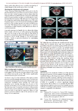
Fig 6 Marking of zones of the prostate
The safety function built into the Sonablate®-500 device
allows you to prevent errors and even interrupt the
procedure if any of the parameters go beyond the safe limits.
The rectal wall is cooled to 16-200 C to prevent tissue
damage. During the entire procedure, the position of the
sensor relative to the rectal wall is constantly monitored.
Measurement of visual visibility and continuous comparison
with reference images (Reflectivity Index Measurment -
RIM). The Sonablate®-500 software allows the surgeon to
adjust the degree of exposure in each of the conditional
zones that were formed during the marking before
performing the ablation. The HIFU signal precisely and
precisely affects the prostate tissue in different zones and
allows you to accurately determine the treatment area in
relation to the borders of the prostate or the external
sphincter of the urinary bladder. After the completion of the
Fig 5 Function of the neurovascular bundle detector. operation, the bladder was drained with a Foley catheter 16-
Wide viewing angle treatment 18 Ch.
A wide viewing angle of 900 allows visual coverage of the
Conclusion
entire gland, and this allows most procedures to be carried HIFU therapy in the treatment of BPH is one of the modern
out without moving the probe. This feature significantly developing minimally invasive methods. Currently, there is
reduces the time spent on treatment. insufficient data on the long-term results of HIFU use in
As shown in fig. 5, for the treatment of the entire prostate patients with BPH. However, with the correct selection of
gland as a whole is divided into several zones. The first zone patients and appropriate indications, the prostate volume is
is always located along the anterior part of the prostate with up to 90 cubic meters. cm, no middle lobe, no complications
subsequent treatment zones towards the rectal wall of BPH, it is possible to achieve significant clinical
(posterior part), thereby ensuring that all parts of the improvement. All this allows us to conclude that the HIFU
prostate are treated. A 4.0 cm probe must be used to treat method has the right to exist as one of the minimally invasive
the front (top row). To treat the central part of the prostate methods of treating prostate adenoma, however, additional
(central row), a 3.0 cm transceiver must be used, if the rectal research is required in this direction.
wall is at least 1.0 cm from the transceiver, then a 4.0 cm Literature
probe is used. the posterior side of the prostate is made [1] Гафаров Р.Р., Аллазов С.А., Гиясов Ш.И. Лазерная
using a 3.0 cm transceiver. энуклеация – новое слово в оперативном лечении
доброкачественной гиперплазии предстательной
железы. Вестник врача 1; 138-144.
[2] Ткачук В.Н., Лукьянов А.Э. Доброкачественная
гиперплазия предстательной железы. - СПб.:
Издательство «Спецлит», 2003.- 130 с.
ID: IJTSRD41125 | Special Issue on Innovative Development of Modern Research Page 145