
Page 149 - Combine
P. 149
International Journal of Trend in Scientific Research and Development (IJTSRD) @ www.ijtsrd.com eISSN: 2456-6470
3. It is necessary to loosen and adjust the position of the
probe so that the transceiver window is at the front and
the reference mark is at zero angular level. After
securing the cuff of the probe manipulator, the probe
must be secured in this position.
4. Next, it is necessary to loosen the central handle of the
probe manipulator and carefully insert the probe tip into
the patient's rectum.
5. When the probe is positioned accurately and satisfactory
preliminary visualization is achieved, the bracket of the
multifunctional probe is rigidly fixed to prevent the
focus of the ablation zone from shifting, after which,
under visual ultrasound guidance in real time, the
prostate is conditionally divided into 3 treatment zones
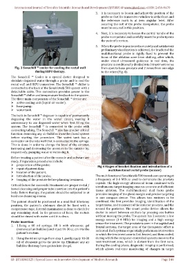
Fig. 3 Sonachill ™ cooler for cooling the rectal wall from apex to base. prostate and 2 zones from one edge
during HIFU therapy. to the other (Fig. 4).
The Sonachill ™ Cooler is a special device designed to
circulate degassed water through a probe and to cool the
rectal wall and HIFU transceiver. The Sonachill ™ chiller is
connected to the back of the Sonablate® 500 system with a
detachable cable. This connection provides power to the
Sonachill ™ chiller and temperature feedback to the system.
The three main components of the Sonachill ™ device are:
active cooling unit (liquid-air cooler);
hose pump;
water tank.
The built-in Sonachill ™ degasser is capable of permanently
degassing the water in the water circuit, making it
unnecessary to use degassed water when first filling the
system. The Sonachill ™ is connected to the probe with
connecting tubing. The Sonachill ™ also has another critical
function: removing any air bubbles from the closed system
before starting the procedure. The water tank has a
connector on the side wall that is connected to the syringe.
This is done in order to change the level of the canister,
increasing and decreasing the pressure in the canister by,
respectively, pumping water or removing it.
Before treating a patient after the console and software are
ready. Preparation procedures include:
preparation of the probe;
Fig 4 Stages of bracket fixation and introduction of a
input of patient data;
multifunctional rectal probe (sensor)
location of the patient;
introduction of the probe; The multifunctional Sonablate®-500 transducer operating at
imaging of the prostate before planning treatment. a frequency of 3-8 MHz is used to demarcate the prostate
capsule. The high-energy ultrasound beam combined with
Critical factors for successful treatment are proper rectal /
simultaneous target imaging ensures accurate and efficient
bowel cleansing and proper tube insertion into the patient's tissue ablation. The multifunctional dual focus probe
body. Before therapy, the patient should be subjected to at
provides imaging of the gland margin and precise targeting
least one cleansing enema.
in one compact device. This allows two emitters to be
The patient should be positioned in a modified lithotomy combined: the first provides imaging, identification of the
position, the patient's abdomen should be fixed with a target tissue, and treatment of the anterior prostate, and the
compression tape. A rectal examination is done to check for second the posterior. The smart probe device allows the
any remaining stool. In the presence of feces, the rectum doctor to select between emitters by pressing one button
should be rinsed with water until it is clean. without moving the probe. The system has 2 sensors: a low
energy sensor (3-4 MHz) for imaging and a high energy
Probe insertion
sensor for treatment. The prostate is seen in the sagittal and
1. 1.Using a 60 ml syringe, fill it with ultrasonic gel
frontal sections, the target area of the therapeutic effect is
(remove all air bubbles) and inject 10-30 cc. cm into the
indicated. Both systems sequentially perform an intervention
patient's rectum.
in which the treatment zone is first heated, and then cooled,
2. Using the 60 ml syringe from step 1, gently apply 10-30 during which the computer-controlled systems move to the
ml of ultrasonic gel to the probe tip. Eliminate any air next treatment zone, which is distant from the first zone.
bubbles that may have gotten into the gel. During the cooling phase, diagnostic imaging is performed,
which allows real-time monitoring of changes in target
ID: IJTSRD41125 | Special Issue on Innovative Development of Modern Research Page 144